


Hidden Realm Of Reef Fish “Rainbow” Fluorescence Hiding In Plain Sight
Recent studies conducted by researchers at the American Museum of Natural History provide insights into the ancient beginnings of biofluorescence in fish and the variety of vibrant colours associated with this biological occurrence. Outlined in two related studies recently released in Nature Communications and PLOS One, the results indicate that biofluorescence has been around for at least 112 million years and has independently evolved over 100 times, primarily among fish inhabiting coral reefs.
The latest research indicates that marine fishes exhibit a wider range of biofluorescent colours than previously documented. Biofluorescence occurs when an organism absorbs light, alters it, and then emits it in a different hue, showcasing various wavelengths of green, yellow, orange, and red.
“Researchers have known for a while that biofluorescence is quite widespread in marine animals, from sea turtles to corals, and especially among fishes,” said Emily Carr, a Ph.D. student in the Museum’s Richard Gilder Graduate School and the lead author on the two new studies. “But to really get to the root of why and how these species use this unique adaptation — whether for camouflage, predation, or reproduction — we need to understand the underlying evolutionary story as well as the scope of biofluorescence as it currently exists.”
In the study published in Nature Communications, Carr conducted an extensive survey of all known biofluorescent teleosts (bony fish that represent the largest group of vertebrates currently in existence). This research identified 459 species that exhibit biofluorescence, including 48 species that were not previously recognised for this trait. The findings suggest that biofluorescence has evolved over 100 times in marine teleosts, with origins tracing back approximately 112 million years, starting with eels. Additionally, the research team discovered that fish species inhabiting or near coral reefs develop biofluorescence at a rate about 10 times higher than those not associated with reefs. There was also a notable increase in the number of fluorescent species following the Cretaceous-Paleogene (K-Pg) extinction event around 66 million years ago, which led to the extinction of all non-avian dinosaurs.
“This trend coincides with the rise of modern coral-dominated reefs and the rapid colonization of reefs by fishes, which occurred following a significant loss of coral diversity in the K-Pg extinction,” Carr said. “These correlations suggest that the emergence of modern coral reefs could have facilitated the diversification of fluorescence in reef-associated teleost fishes.”
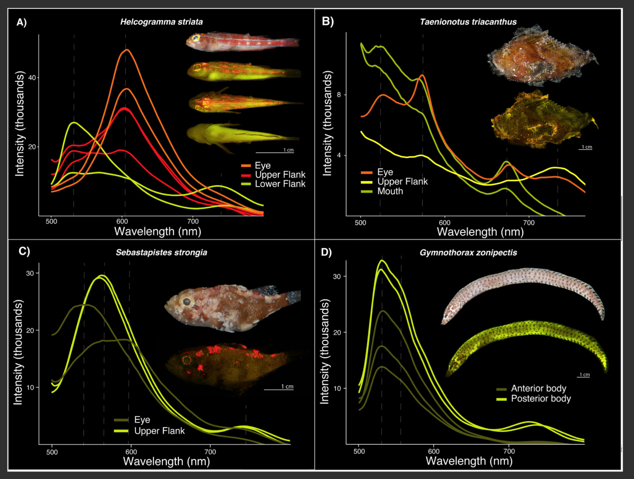
Among the 459 biofluorescent teleosts identified in this study, most are linked to coral reefs. In the PLOS One research, Carr and his team utilised a unique photography setup featuring ultraviolet and blue excitation lights along with emission filters to examine the light wavelengths emitted by fishes in the Museum’s Ichthyology collection. These specimens, gathered over the past fifteen years during Museum expeditions to locations such as the Solomon Islands, Greenland, and Thailand, had previously shown signs of fluorescence, but the complete spectrum of their biofluorescent emissions remained a mystery. This new research uncovers a much greater variety of colours emitted by teleosts — with some families displaying at least six different fluorescent emission peaks that align with wavelengths spanning several colours — than what was known before.
“The remarkable variation we observed across a wide array of these fluorescent fishes could mean that these animals use incredibly diverse and elaborate signalling systems based on species-specific fluorescent emission patterns,” said Museum Curator John Sparks, an author on the new studies and Carr’s advisor. “As these studies show, biofluorescence is both pervasive and incredibly phenotypically variable among marine fishes. What we would really like to understand better is how fluorescence functions in these highly variable marine lineages, as well as its role in diversification.”
The researchers point out that the various wavelengths of fluorescent emissions discovered in this study may have significant implications for the identification of new fluorescent molecules, which are commonly utilised in biomedical fields, such as fluorescence-guided disease diagnosis and treatment. The other contributors to this research include Rene Martin from the Museum and the University of Nebraska-Lincoln; Mason Thurman from Clemson University; Karly Cohen from California State University; Jonathan Huie from George Washington University; David Gruber from Baruch College and The Graduate Center, City University of New York; and Tate Sparks from Rutgers University.
[Image: Fig 5. Variation in fluorescent emission spectra, or lack thereof, over different regions of the body in individuals. A) Helcogramma striata (Tripterygiidae, n = 1); B) Taenianotus triacanthus (Scorpaenidae, n = 1); C) Sebastapistes strongia (Scorpaenidae, n = 1); D) Gymnothorax zonipectis (Muraenidae, n = 2). Note: For each panel the top image shows the species imaged under white light and all others show the species fluorescing. Dashed lines represent fluorescent emission peaks. Intensity values are relative and do not signify overall brightness]
Emily M. Carr, Mason A. Thurman, Rene P. Martin, Tate S. Sparks, John S. Sparks. Marine fishes exhibit exceptional variation in biofluorescent emission spectra. PLOS One, 2025; 20 (6): e0316789 DOI: 10.1371/journal.pone.0316789


